CPDA-1- anticoagulant citrate phosphate dextrose adenine solution
CPDA-1 by
Drug Labeling and Warnings
CPDA-1 by is a Prescription medication manufactured, distributed, or labeled by Fenwal, Inc., Fenwal International, Inc.. Drug facts, warnings, and ingredients follow.
Drug Details [pdf]
-
Instructions for Blood Collection Using Anticoagulant Citrate Phosphate Dextrose Adenine Solution, USP (CPDA-1) BLOOD-PACK™ Units with Elongated Primary Container
Rx only
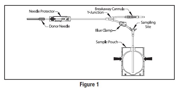
Contains Sample Diversion System for the collection of unanticoagulated whole blood samples for laboratory testing and the DONORCARE™ Needle Guard. The elongated primary container may be used to collect and store 450 mL of blood for 35 (CPDA-1) days. It may also be used to prepare and store frozen red cells using the high glycerol (40% W/V) technique.
Use aseptic technique.
Notes:
- If Sample Diversion System is not used, donor samples may be collected using an alternate method following standard procedures.
- Nominal tubing dimensions of product are 0.118" inner diameter x 0.025" wall thickness.
Precautions:
- Upon removal of BLOOD-PACK™ unit from the clear plastic overwrap, visually inspect the unit.
- Do not use the product if the in-line cannula is broken and/or anticoagulant is present in the sample pouch or in the tubing from the in-line cannula to the sample pouch and donor needle (see Figure 1). Note that condensation in the empty tubing of the BLOOD-PACK unit is expected as a result of the sterilization process.
- Do not use unless the solutions are clear.
- Since plastics become brittle at freezer temperatures, use caution during handling to prevent container breakage.
- The elongated primary container should not be used for the storage of red cells at liquid nitrogen temperatures.
- 1. Identify BLOOD-PACK unit using appropriate donor identification system.
- 2. Adjust donor scale to desired collection weight.
- 3. Position primary container on the donor scale as far as possible below donor arm.
- 4. Clamp donor tubing between the needle and Y-junction with hemostat. (This step can be performed prior to step 1 or 2.)
- 5. Visually inspect the tubing from the in-line cannula to the sample pouch and donor needle, as well as the sample pouch to reconfirm that there is no anticoagulant present.
Note: Ensure that the sample pouch remains below the donor’s arm.- 6. Apply pressure to donor’s arm and disinfect site of venipuncture.
- 7. If blood pressure cuff is used, inflate to approximately 60 mmHg.
- 8. Remove needle cover per instructions below:
- a) Holding the hub and cover near the tamper-evident seal, twist cover 1/4 turn to break seal.
- b) Remove needle cover, being careful not to drag the cover across the needle point.
- 9. Perform venipuncture, appropriately secure donor needle and/or tubing and release hemostat.
- 10. When good blood flow is established, slide the DONORCARE™ needle guard over the needle hub into the engaged position. Leave the front third of the needle hub exposed for access. Stabilize the front of the needle guard to arm with tape. (see Figure 2)
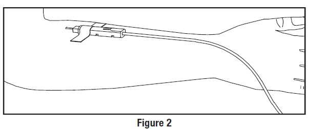
Note: In difficult collection conditions (e.g. slow blood flow), leave the needle guard disengaged behind the hub during collection.
Engage the needle guard at the end of blood collection.- 11. Allow the sample pouch to fill with blood according to center procedure. Monitor blood flow into sample pouch.
Notes:
- The sample pouch contains an average fill volume of approximately 53 mL with a maximum fill volume of approximately 60 mL when filled to capacity.
- If less blood sample volume is required, the flow to the sample pouch may be stopped prior to completely filling the pouch. For example, in order to target a fill volume of approximately 40 mL, fill to the level indicated by the arrows in Figure 1. Ensure the pouch is hanging vertically.
- The tube leading from the Y-junction to the sample pouch contains an additional volume of approximately 2 mL.
Precautions:
- Do not elevate or squeeze the sample pouch as this could cause blood to backflow from the sample pouch into the collection system.
- Once the sample pouch is filled to desired volume, complete steps 12 - 20 within approximately 4 minutes to avoid possible clot formation in the tubing and/or sample pouch.
- 12. Close the blue clamp on tubing between the Y-junction and the sample pouch.
- 13. Break the in-line cannula below the Y-junction in the donor tubing to the primary container allowing blood collection to proceed. To completely break the in-line cannula, grasp with both hands. Snap it at a 90° angle in one direction, and then bend it at a 90° angle in the opposite direction. Ensure the in-line cannula is completely broken and that the blood flows freely to the primary container.
Precaution: Failure to break the in-line cannula completely may result in restricted blood flow.
- 14. Mix blood and anticoagulant in primary container at several intervals during collection and immediately after collection.
- 15. Following blood center procedures, hermetically seal the tubing between the sampling site and the Y-junction to maintain sterility of the blood collection system prior to removing blood samples.
Warning:
- Do not proceed with the remaining steps until the tubing leading to the sample pouch is hermetically sealed between the sampling site and the Y-junction. To maintain the whole blood collection container as a closed system, the tubing between the sample pouch and Y-junction must be hermetically sealed prior to inserting the access device into the sampling site. Failure to do so may lead to contamination of the whole blood collection.
- 16. Insert the access device by pushing firmly into the sampling site until the membrane seal is penetrated.
Note: If the access device is assembled such that the outer barrel is screwed onto the Luer, make sure to rotate clockwise upon insertion to avoid barrel detaching from Luer.
- 17. Open the cap on the access device (if applicable). Hold access device so that the sample pouch hangs down.
- 18. Directly align the vacuum sample tube with the internal needle in the access device. Insert vacuum sample tube into device.
- 19. Allow vacuum sample tube to fill with blood then remove from the access device.
- 20. Repeat steps 18 and 19 until the desired number of vacuum sample tubes have been filled.
Notes:
- If the access device needs to be replaced, use a hemostat to clamp the tubing between the sampling site and the sample pouch. Then, grasp base of sampling site with one hand and pull the access device out with the other hand. Firmly insert the new access device. Remove hemostat and continue sampling.
- If the access device is assembled such that the outer barrel is screwed onto the Luer, make sure to rotate clockwise upon removal to avoid barrel detaching from Luer.
- The access device can only be replaced one time.
Precaution: When replacing access device, be careful to avoid contact with any blood droplets on the Luer or sampling site. Discard used access device appropriately.
- 21. Collect only 450 ± 45 mL of blood into the elongated primary container.
Precaution: The primary container of this blood collection system is larger than conventional primary containers and will not appear as full as smaller containers when 450 mL of blood has been collected. The amount of blood collected should be determined by weight for collection of 450 mL of blood.Precaution: Once the desired blood volume is collected, complete steps 22-25 within approximately 4 minutes to avoid possible clot formation in the tubing.
- 22. Release pressure on the donor’s arm. If appropriate, apply hemostat to donor tubing between the needle and the Y-junction.
- 23. Hermetically seal donor tubing near in-line cannula on side leading to the primary container.
- 24. Withdrawal of Needle (see Figure 3)
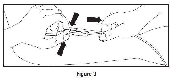
Precaution: The needle guard must be held stationary while the needle is withdrawn into it.
- a) Place folded sterile gauze over puncture site and hold in place with finger tip without exerting pressure.
- b) Hold sides of needle guard near the front, between the index finger and thumb. Pull the tubing smoothly until the needle is locked into the needle guard.
- c) Confirm the needle lock by:
- Listen for the 2nd “click” as the needle is drawn into the needle guard.
- Ensure the tubing cannot be pulled through the needle guard.
- 25. Strip blood from donor tubing into primary container, mix and allow the tubing to refill; repeat once.
- 26. Seal at X marks on donor tubing to provide numbered aliquots of anticoagulated blood for typing or crossmatching.
Note: Step 27 may be performed prior to step 25 or 26 if desired.
- 27. Remove and discard the Sample Diversion System and needle guard into an appropriate biohazardous waste container following established procedures.
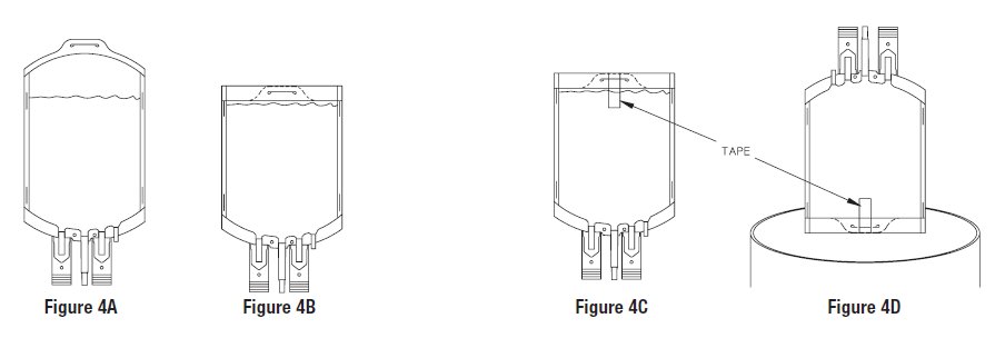
- 28. For component preparation, prior to centrifugation, the excess material at bottom of the elongated primary container should be rolled over as follows:
- To perform bottom rolling, invert primary container (Figure 4A).
- With ports down, roll over excess container material (Figure 4B) and secure with tape (Figure 4C).
- Place the rolled primary container in an upright position (ports up) on top of secondary containers in centrifuge cup (Figure 4D).
- 29. When processing a multiple BLOOD-PACK unit, centrifuge primary and secondary containers to prepare CPDA-1 Red Blood Cells.
- 30. Gently remove the (bottom-rolled) elongated primary container from centrifuge cup. Using care to ensure that bottom fold area is supported, place the container in the plasma extractor.
- 31. Express plasma into empty TRANSFER PACK™ container by releasing pressure plate and opening closure in tubing of primary container. All visible (platelet rich) plasma should be removed if cells are to be frozen subsequently.
- 32. When desired amount of plasma has been removed, clamp tubing between Y and plasma container, remove tape, and unfold unit.
- 33. Seal transfer tubing in three places between the Y-connectors. Cut middle seal being careful to avoid fluid splatter.
- 34. For further processing with multiple BLOOD-PACK units, use standard component processing and storage techniques.
For Liquid Red Cell Storage:
- 35. Store suspended CPDA-1 Whole Blood/Red Blood Cells between 1 and 6°C and infuse within 35 days of collection.
For Frozen Red Cell Storage:
Precaution: Freeze within 6 days of collection. Use aseptic technique during red cell glycerolization.
- 36. If remaining Red Blood Cells are intended for frozen storage in the elongated primary container, the hematocrit should be between 75 and 80% and one empty container should be left attached to the primary for glycerol removal.
- 37. Supplies Required
- A) GLYCEROLYTE™ 57 Solution, 500 mL (Code 4A7833)
- B) Plasma Transfer Set (Code 4C2243)
- C) Sterile Filtered Airway Needle
- D) Labels for elongated primary freezing pack and cardboard storage container
- E) 1 heat-sealable 8" x 12" plastic bag (available from 3M Company, St. Paul, Minnesota)
- F) Corrugated 7" x 5" x 1-1/2" cardboard protective container (available from NABI Company, Woburn, Massachusetts)
- G) 70% isopropyl alcohol swabs
- H) Waterproof tape (available from 3M Company, St. Paul, Minnesota)
- I) Hand sealer clips (Code 4R4418)
- J) Agitator
- K) Plasma Extractor (Code 4R4414)
- 38. Allow glycerol solution and red cells to warm to 22-30°C prior to adding solution to red cells. Process cells within 4 hours after removal from refrigeration.
- 39. Remove all but one numbered tubing segment. Heat seal the remaining segment as close to the container as possible. Leave the segment attached to the container.
- 40. Weigh the unit of red cell concentrate and subtract the tare weight (approx. 72 grams for the elongated primary container and integrally attached TRANSFER PACK container). The net weight of the red cells should not exceed 330 grams.
- 41. Place the red cell concentrate on a platform agitator.
- 42. Remove the metal tab from the top of the glycerol solution bottle, disinfect the stopper with 70% isopropyl alcohol and aseptically insert one of the plasma transfer set couplers into the bottle outlet port. Close roller clamp on plasma transfer set.
- 43. Insert the filtered airway needle into the bottle vent port.
- 44. Invert the bottle and suspend it 18 inches above the level of the primary bag on the agitator.
- 45. Aseptically insert the remaining transfer set coupler of the transfer set into the delivery tube assembly on the tubing leading from the elongated primary container.
- 46. Glycerol solution should be added to cells based on cell volume and in three steps. Refer to Table 1.
Table 1 - Method of Addition of GLYCEROLYTE 57 Solution to Red Cell Concentrates
Gross Weight
of Unit (g)*
Net Weight
of Unit (g)*
Initial Addition
of Glycerol (mL)
Second Addition
of Glycerol (mL)
Third Addition
of Glycerol (mL)
Total Glycerol
Added (mL)
218-272
150-200
50
50
250
350
273-312
201-240
50
50
350
450
313-402
241-330
50
50
400
500
*Tare weight of the empty elongated primary container and integrally attached TRANSFER PACK container is approximately 72 grams.
- 47. Adjust platform agitator to 180 oscillations/minute. Occlude tubing between elongated primary collection container and empty TRANSFER PACK container by folding tubing and placing a hand sealer clip over fold (do not crimp) to prevent flow of solution into TRANSFER PACK container.
- 48. Add 50 mL of glycerol solution to the red cells while the cells are being agitated.
- 49. Turn off agitator and allow equilibration for 5 minutes.
- 50. Restart agitator; add 50 mL of glycerol solution to cells.
- 51. Turn off agitator and allow equilibration for 2 minutes.
- 52. Remove red cells from agitator.
- 53. Refer to Table 1 to determine remaining volume of glycerol solution to be added to red cells. Use continuous, vigorous manual agitation during solution addition.
- 54. Seal tubing proximal to the delivery tube assembly. Ensure that the TRANSFER PACK container remains integrally attached to the primary collection bag. Discard the glycerol bottle, connector tubing, and delivery tube assembly.
- 55. Centrifuge the glycerolized red cells at 1250 x g for 10 minutes.
- 56. Suspend primary container in plasma extractor, remove hand sealer clip, and express all visible supernatant glycerol from the red cell container into the TRANSFER PACK container.
- 57. Seal the tubing 2 inches from the red cell container, separate tubing and discard the TRANSFER PACK container containing the supernatant glycerol solution.
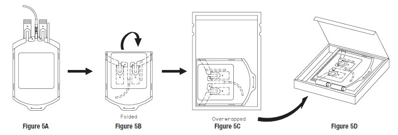
- 58. Appropriately label product (Figure 5A).
- 59. Fold over the top portion of the primary bag approximately 2 inches (Figure 5B), place the unit into the 8" x 12" plastic overwrap (Figure 5C) and seal across the top using an impulse sealer so that there is as little trapped air as possible. Make sure that the ports and tubing segments are folded beneath the unit so that they are protected from breakage when frozen.
- 60. Place the glycerolized red cells and overwrap into the cardboard box (Figure 5D). Close the box and seal the flaps with tape. Label the box, and place in the bottom of a -80°C freezer. To facilitate freezing, do not stack units. After 24 hours, the frozen units can be stacked and/or stored in other -80°C freezers.
- 61. After storage, thaw and deglycerolize red cells according to standard institutional procedure.
- 62. Infuse deglycerolized cells within 24 hours after processing.
References
Freezing in the Primary Polyvinylchloride Plastic Collection Bag: A new system for freezing nonrejuvenated and rejuvenated Red Blood Cells. C.R. Valeri, D.A. Valeri, J. Anastasi, J.J. Vecchione, R.C. Dennis, and C.P. Emerson. Transfusion 1981; 21:138-149.
 Manufacturer
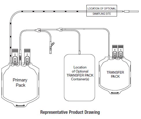
FENWAL, BLOOD-PACK, GLYCEROLYTE, and TRANSFER PACK are trademarks of Fenwal, Inc.
DONORCARE is a trademark of ITL Corporation.
© 2010 Fenwal, Inc. All rights reserved.
Store at Controlled Room Temperature.
USP Definition of “Controlled Room Temperature”
United States Pharmacopeia, General Notices.
United States Pharmacopeial Convention, Inc.
12601 Twinbrook Parkway, Rockville, MD Fenwal, Inc.
Lake Zurich, IL 60047 USAMade in USA
1-800-933-6925
07-19-04-834 REV: A
05/2010 -
PACKAGE/LABEL DISPLAY PANEL
Code 4R1250
3 Units
Fenwal™
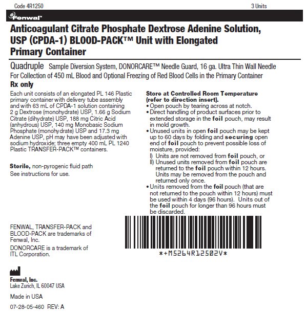
Anticoagulant Citrate Phosphate Dextrose Adenine Solution, USP (CPDA-1) BLOOD-PACK™ Unit with Elongated Primary Container
Quadruple Sample Diversion System, DONORCARE™ Needle Guard, 16 ga. Ultra Thin Wall Needle
For Collection of 450 mL Blood and Optional Freezing of Red Blood Cells in the Primary ContainerRx only
Each unit consists of an elongated PL 146 Plastic primary container with delivery tube assembly and with 63 mL of CPDA-1 solution containing 2 g Dextrose (monohydrate) USP, 1.66 g Sodium Citrate (dihydrate) USP, 188 mg Citric Acid (anhydrous) USP, 140 mg Monobasic Sodium Phosphate (monohydrate) USP and 17.3 mg Adenine USP, pH may have been adjusted with sodium hydroxide; three empty 400 mL PL 1240 Plastic TRANSFER-PACK™ containers.
Sterile, non-pyrogenic fluid path
See instructions for use.Store at Controlled Room Temperature (refer to direction insert).
- Open pouch by tearing across at notch.
- Direct handling of product surfaces prior to extended storage in the foil pouch, may result in mold growth.
- Unused units in open foil pouch may be kept up to 60 days by folding and securing open end of foil pouch to prevent possible loss of moisture, provided:
- I) Units are not removed from foil pouch, or
- II) Unused units removed from foil pouch are returned to the foil pouch within 12 hours. Units may be removed from the pouch and returned only once.
- Units removed from the foil pouch (that are not returned to the pouch within 12 hours) must be used within 4 days (96 hours). Units out of the foil pouch for longer than 96 hours must be discarded.
FENWAL, TRANSFER-PACK and BLOOD-PACK are trademarks of Fenwal, Inc.
DONORCARE is a trademark of ITL Corporation. Fenwal, Inc.
Lake Zurich, IL 60047 USAMade in USA
07-28-05-460 REV: A
-
INGREDIENTS AND APPEARANCE
CPDA-1
anticoagulant citrate phosphate dextrose adenine solutionProduct Information Product Type HUMAN PRESCRIPTION DRUG Item Code (Source) NDC: 0942-6322 Route of Administration INTRAVENOUS Active Ingredient/Active Moiety Ingredient Name Basis of Strength Strength Dextrose Monohydrate (UNII: LX22YL083G) (ANHYDROUS DEXTROSE - UNII:5SL0G7R0OK) Dextrose Monohydrate 2 g in 63 mL Trisodium Citrate Dihydrate (UNII: B22547B95K) (Anhydrous Citric Acid - UNII:XF417D3PSL) Anhydrous Citric Acid 1.66 g in 63 mL Anhydrous Citric Acid (UNII: XF417D3PSL) (Anhydrous Citric Acid - UNII:XF417D3PSL) Anhydrous Citric Acid 188 mg in 63 mL Sodium Phosphate, Monobasic, Monohydrate (UNII: 593YOG76RN) (PHOSPHATE ION - UNII:NK08V8K8HR) Sodium Phosphate, Monobasic, Monohydrate 140 mg in 63 mL Adenine (UNII: JAC85A2161) (Adenine - UNII:JAC85A2161) Adenine 17.3 mg in 63 mL Inactive Ingredients Ingredient Name Strength Sodium Hydroxide (UNII: 55X04QC32I) Packaging # Item Code Package Description Marketing Start Date Marketing End Date 1 NDC: 0942-6322-04 63 mL in 1 BAG; Type 0: Not a Combination Product Marketing Information Marketing Category Application Number or Monograph Citation Marketing Start Date Marketing End Date NDA BN770420 06/24/2007 Labeler - Fenwal, Inc. (794519020) Establishment Name Address ID/FEI Business Operations Fenwal International, Inc. 091164590 MANUFACTURE(0942-6322)
© 2026 FDA.report
This site is not affiliated with or endorsed by the FDA.